

Radiological features:
- Abdominal X-Ray: Look for a calcified appendicolith in the right lower quadrant (RLQ). Other indicators include free air; small bowel ileus; extra-luminal gas; caecal wall thickening; loss of pelvis fat planes around the bladder suggests pelvic free fluid; loss of the properitoneal fat line; psoas line distortion and abrupt cut-off of the normal gaseous pattern at the hepatic flexure due to colonic spasm.
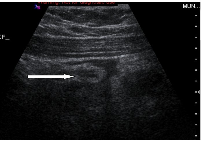
- Ultrasonography: Suggestive features include an obstructing appendicolith – a blind ending non-peristaltic, non-compressible tubular structure and prominent vasculature within the meso-appendix; wall thickness should be 2mm in a normal appendix or 6mm in total diameter.
 |
| Large calcified appendicolith (arrowhead) |
 |
| Inflammed appendix displaying thick wall |
- CT: Sensitive and specific investigation. Not routine due to radiation dose. Luminal distension with a thickened enhancing wall (+/-) an appendicolith. Local inflammation shows as linear streaking in the adjacent fat. Abscesses may be present.
- Contrast investigations: Occasionally picked up coincidently. Suggested by non-filling or localised mucosal oedema within the caecal pole.
 |
| Inflammed appendix with multiple appendicoliths |
 |
| Normal appendix; barium enema radiographic examination |
No comments:
Post a Comment